
“I have specialised in cellulite and skin tightening for 25+ years, in post-liposuction fibrosis for 20+ years; and in skin ultrasonography for cellulite, skin laxity, lipedema and fibrosis for 4 years now. During this time I have performed hundreds of scans, so I can help you understand your skin better and offer you the best possible treatment with the aid of ultrasound skin imaging.”
Cellulite Ultrasonography™: a LipoTherapeia exclusive
Assess and better understand your post-liposuction fibrosis and adhesions, and more
Get answers to your key questions about your cellulite, lipedema, skin laxity, fibrosis and fat
High-tech equipment and clear, science-based explanations in plain language
Use Cellulite Ultrasonography™ to make the most of our treatments
The only aesthetic practice in London specialising 100% in skin tightening & cellulite removal
Book an expert cellulite / skin tightening treatment at our central London clinic
More ultrasound skin images from the same client:
Diagnostic ultrasound images of skin at the lower back of thighs, with loose skin, intermediate fascia and large cellulite “bump” visible
Diagnostic ultrasound images of skin at the lower buttocks, with skin laxity
Diagnostic ultrasound images of skin at the mid-upper buttocks, with cellulite
Expert ultrasound skin imaging of cellulite, skin laxity, fat tissue, post-liposuction fibrosis, lipedema and more at LipoTherapeia
Cellulite Ultrasonography™ | Exclusively at LipoTherapeia®, London
We are proud to be the first and only cellulite clinic in the world to offer Cellulite Ultrasonography™ to our clients (since 2022):
For accurate assessment and enhanced results with our cellulite treatments
To help people understand and get answers to their questions about fibrosis, scar tissue and adhesions that commonly occur after liposuction, other cosmetic surgery, non-surgical cosmetic treatments and even injuries that can sometimes leave skin with scar tissue, dents and bumps
And also to help people understand and get answers to their questions about lipedema versus: normal female adiposity, cellulite, lipedema, water retention and skin laxity
⏵ Check prices and book an expert skin ultrasound scan at our London clinic
Look inside your skin™ and assess your cellulite; differentiate it from lipedema, fat or other issues; and better understand your skin, fascia and fat
With our skin ultrasonography service you can assess, differentiate and better understand your specific case, by seeing how your skin looks from the inside and make informed decisions, in regard to:
Cellulite
Skin laxity
Lipedema
Subcutaneous fat levels
Subcutaneous fascia layers
⏵ Check prices and book an expert skin ultrasound scan at our London clinic
Assess and better understand your post-liposuction fibrosis and adhesions, and more
You can also assess the presence and severity of fibrosis, scar tissue and adhesions after:
Liposuction, vaser, smart lipo, bodytite, airsculpt and more
Abdominoplasty / tummy tuck, thigh lift, arm lift and more
Brazilian butt lift / BBL
Cellulite surgery, such as cellulaze, cellutite, cellfina, subcision, aveli and more
Non-surgical - but harsh for the skin and underlying tissues - procedures, such as RF microneedling, extreme intensity HIFU, extreme intensity RF, extreme intensity ultrasound etc
Hyaluronic acid fillers or poly-L-lactic acid collagen-stimulating injectables, such as Sculptra
Previous mechanical previous injuries that may sometimes leave skin with scar tissue, adhesions, deep fibrosis, dents and bumps
⏵ Check prices and book an expert skin ultrasound scan at our London clinic
Get answers to your key questions about your cellulite, lipedema, skin laxity, fibrosis and fat
Our skin ultrasonography scan and consultation can offer you answers to the following common questions, among many others:
Is what I have cellulite, plain fat, skin looseness, lipedema, visceral fat or water retention - and how can it be improved?
I had liposuction / vaser / smart lipo / cellulite surgery, can you check if what I have is fibrosis or unremoved fat?
My skin is damaged due to RF microneedling / HIFU / extreme intensity RF / extreme intensity ultrasound / poly-L-lactic acid fillers, what can I do?
Why is my skin so loose and where to focus to make it tighter?
⏵ Check prices and book an expert skin ultrasound scan at our London clinic
High-tech equipment and clear, science-based explanations in plain language
At LipoTherapeia we use the latest technology in high-resolution, high-frequency diagnostic ultrasound, specific to skin and subcutaneous adipose tissue. This enables us to clearly visualise skin laxity, cellulite, fat accumulation, lipedema and fibrosis.
Everything will be shown and explained to you clearly in simple terms and you can see and understand how the different structures inside your skin affect your appearance.
If there is scar tissue / fibrosis, those will be clearly seen and we can differentiate between fibrosis, water retention, fat, cellulite and skin laxity, both deep and superficial.
(DISCLAIMER: Please note that this service is for your informational purposes only and does not constitute medical diagnosis, advice or treatment.)
⏵ Check prices and book an expert skin ultrasound scan at our London clinic
Use Cellulite Ultrasonography™ to make the most of our treatments
The 45’ appointment includes full assessment and consultation and optional treatment plan for cellulite reduction and/or skin tightening.
If you wish to have treatment with us, the ultrasound scan, assessment and consultation will be used to add to the visual assessment and optimise your course of treatments and maximise your results.
⏵ Check prices and book an expert skin ultrasound scan at our London clinic
Hundreds of cases over the last four years
Since 2022 we have performed hundreds of scans and have accumulated specialist experience in this subject, which helps us assist our clients with better treatment and better understanding of their skin and underlying tissues.
⏵ Check prices and book an expert skin ultrasound scan at our London clinic
View scan and assessment examples from real clients, below
Below you can see an extensive range of scans from a real client, with full descriptions and explanations.
This is the level of service you will receive.
⏵ Check prices and book an expert skin ultrasound scan at our London clinic
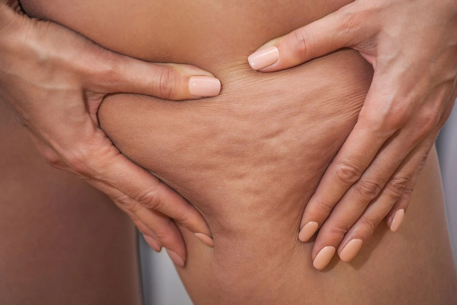
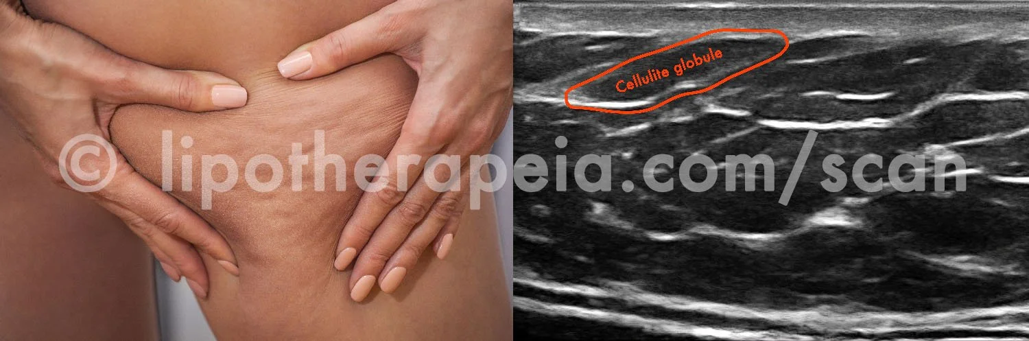
Diagnostic ultrasound image and video of cellulite (hypodermis) and fat tissue (subcutaneous adipose tissue) on the back of thighs
Below you can see ultrasound imaging of a small sized banana roll (upper back of thighs), from a normal body sized woman in her early 40s, with progressed skin laxity and Grade 2 cellulite.
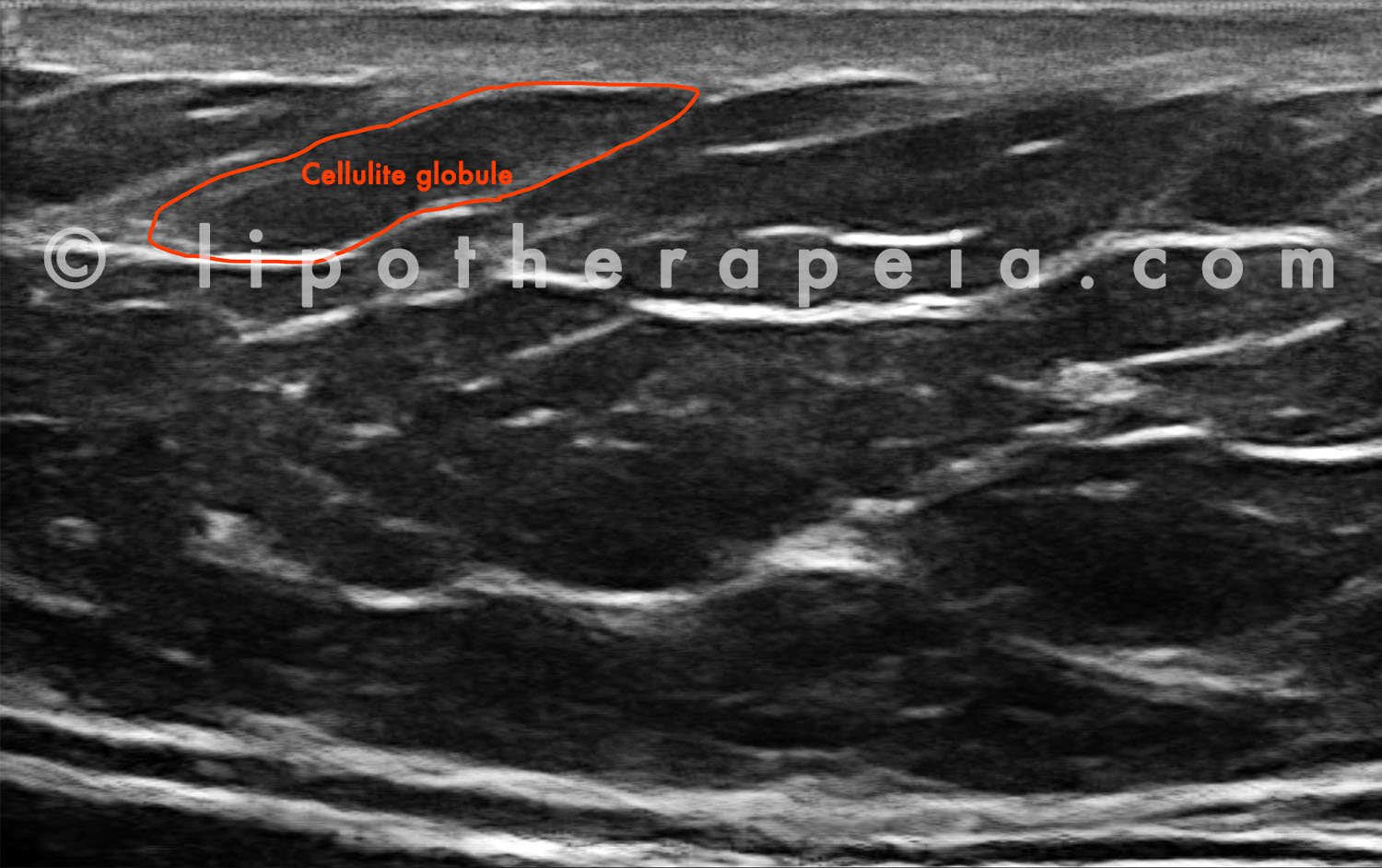
Skin ultrasound image of cellulite globules and subcutaneous fat in the banana roll area, below the buttocks
Skin ultrasound video of cellulite globules and subcutaneous fat in the banana roll area, below the buttocks
In the image and video above, the epidermis, dermis, dermal hypodermal junction / DHDJ, hypodermis, cellulite, cellulite globules, cutaneous retinaculae (“collagen bands” / septae), superficial fascia, subcutaneous adipose tissue (‘fat’), deep fascia, perimuscular fascia and a small part of musculature can clearly be seen.
In the video the alternating cellulite fat globules are visualised even more clearly.
Findings:
The dermis is of normal thickness for that body area
As this banana roll is small-sized, the subcutaneous fat is relatively thin here
The cellulite globules are of average/large size
The intermediate fascia is fragmented
The tissue is quite fibrous, which is normal for this body area
These findings, together with those of other body areas, the external assessment and information provided during the consultation, were used to optimise treatment for this client.
All images were taken at our clinic in London (LipoTherapeia, 10 Portman Square, London W1).
At the time of first publishing this page the client was currently undergoing treatment for both cellulite and skin laxity with deep-acting, high-power radiofrequency and high-power ultrasound cavitation.
Images U1 (C10), U1 (C9)
You can view more images of cellulite from different body areas here
You can additionally view images of skin laxity on different body areas here
You can also view images of fibrosis after cosmetic surgery and unsafe cosmetic procedures here
⏵ Check prices and book an expert skin ultrasound scan at our London clinic
Advanced cellulite and skin tightening treatments in London, by LipoTherapeia
Our cellulite and skin tightening treatments are based on LipoTherapeia® Plus, the strongest SAFE cellulite / skin tightening technology combination available today, skilfully applied without compromises or cut corners, for maximum results, naturally.
LipoTherapeia® Plus comprises 5 exclusive elements:
Deep-acting, high-power radiofrequency, the most effective SAFE cellulite reduction technology, with the best equipment currently available
Deep-acting, high-power ultrasound cavitation, the second most effective SAFE cellulite reduction technology, with the best equipment currently available
High-power LED phototherapy, to maximise and enhance the effects of our high-power, deep-acting radiofrequency and ultrasound, with the best equipment currently available
Advanced treatment protocols, based on actual Physics and the Anatomy & Physiology of cellulite, in order to make the most of these technologies (this is in contrast to the usual one-day training on some basics of how to use the machine, which is the norm at almost all salons and clinics that offer cellulite treatments - including cosmetic surgery practises)
Treatment based on more than two decades of experience, study and research in cellulite and skin tightening, with more than 23,000+ treatments and 4,100+ cases (including 14+ years and 13,000+ high-power, deep-acting radiofrequency and cavitation treatments)
A PAINLESS, realistically priced and effective technology, LipoTherapeia® Plus has a 99.5%+ safety rate at our clinic, as it is based on naturally stimulating collagen production, hypodermal fat release and circulation/lymphatic drainage - not on burning the skin inside, as some procedures mentioned above aim.
In addition to cellulite reduction, the technologies comprising LipoTherapeia® Plus also have a profound effect on skin tightness* as well as a beneficial effect on stretch marks. So you get a combined benefit by the use of these technologies.
(*In fact, deep-acting, high-power radiofrequency is the gold standard technology for SAFE and effective skin tightening.)
In most cases of cellulite / skin laxity we recommend 6-12 sessions for best results (there are no miracle treatments that reduce cellulite or tighten skin in 1-4 sessions).
⏵ Book an expert cellulite / skin tightening treatment at our central London clinic
The only aesthetic practice in London that specialises 100% in skin tightening & cellulite removal
Most aesthetic clinics offer 20, 30, even 50 different treatments, from laser hair removal to fillers to chemical peels to cellulite, and thereby lose specialisation and expertise.
A jack of all trades therapist or clinic cannot possibly be experts in 20, 30 or 50 different treatments. It is physically impossible to follow the science and all the latest developments in 50, 30 or even 20 fields.
And it is impossible to be an expert in an aesthetic condition, if you never study and practise with focus and depth the science of that aesthetic condition.
So at LipoTherapeia we only focus 100% on cellulite reduction and skin tightening - nothing else.
We do not do botox, fillers, chemical peels, dermablading, microdermabrasion, waxing, nails, fluffy facials, laser hair removal and the like: just cellulite reduction and skin tightening with the best ultrasound, radiofrequency and LED phototherapy technologies in the world (no exaggeration). And we have a deep knowledge of these five subjects - for your benefit.
⏵ Book an expert cellulite / skin tightening treatment at our central London clinic
More than two decades of experience in cellulite reduction
At LipoTherapeia we make the most out of the best technologies in the world with our advanced protocols, developed over more than two decades and based on:
A deep knowledge of the Physics of radiofrequency, ultrasound and phototherapy and the anatomy of cellulite and connective tissue (as opposed to the usual one-day training provided by machine manufacturers)
Literally tens of thousands of sessions on thousands of clients with hundreds of kinds of body types, sensitivities and health/aesthetic issues
All with 99.5%+ safety and NO PAIN, NO DOWNTIME, no microneedling, no injections, no fillers, no unsafe “instant miracle treatments”, no numbing creams, no invasiveness, no surgery, no drama.
Just comfortable, safe and effective treatment that works by stimulating your skin to become smoother and firmer., naturally and safely.
Please note: All consultations and treatments are currently provided by cellulite / skin tightening specialist Georgios Tzenichristos, to ensure high quality treatment. If, for religious or other reasons, you wish to have a female therapist or have a female chaperone present, unfortunately we will not be able to assist you, so in this case kindly do not book any sessions with us.
⏵ Book an expert cellulite / skin tightening treatment at our central London clinic
Caring, honest, boutique treatment and advice with an expert - premium treatment without the premium prices
Treatment is always provided by a highly experienced therapist and is personalised, according to your specific needs, after an extensive assessment of your type of cellulite.
No treatment by machine operators trained for one day, no ever-changing therapists and receptionists, no armies of pushy salespeople, no hard sales and no premium prices.
Our focus is on honest, realistic, science-based treatment, combined with caring, professional service, with a smile.
We will be pleased to see you, assess your cellulite or skin laxity, listen to your story, discuss your case and offer you the best possible treatment.
⏵ Book an expert cellulite / skin tightening treatment at our central London clinic
More ultrasound skin images from the same client
Here you can see more images from the same client to help you understand some of the different anatomical regions and what they reveal.
Again, don’t worry about the technical details. The scan results and assessment will be communicated to you in simple and practical terms.
Diagnostic ultrasound images of skin at the lower back of thighs, with loose skin, intermediate fascia and large cellulite “bump” visible
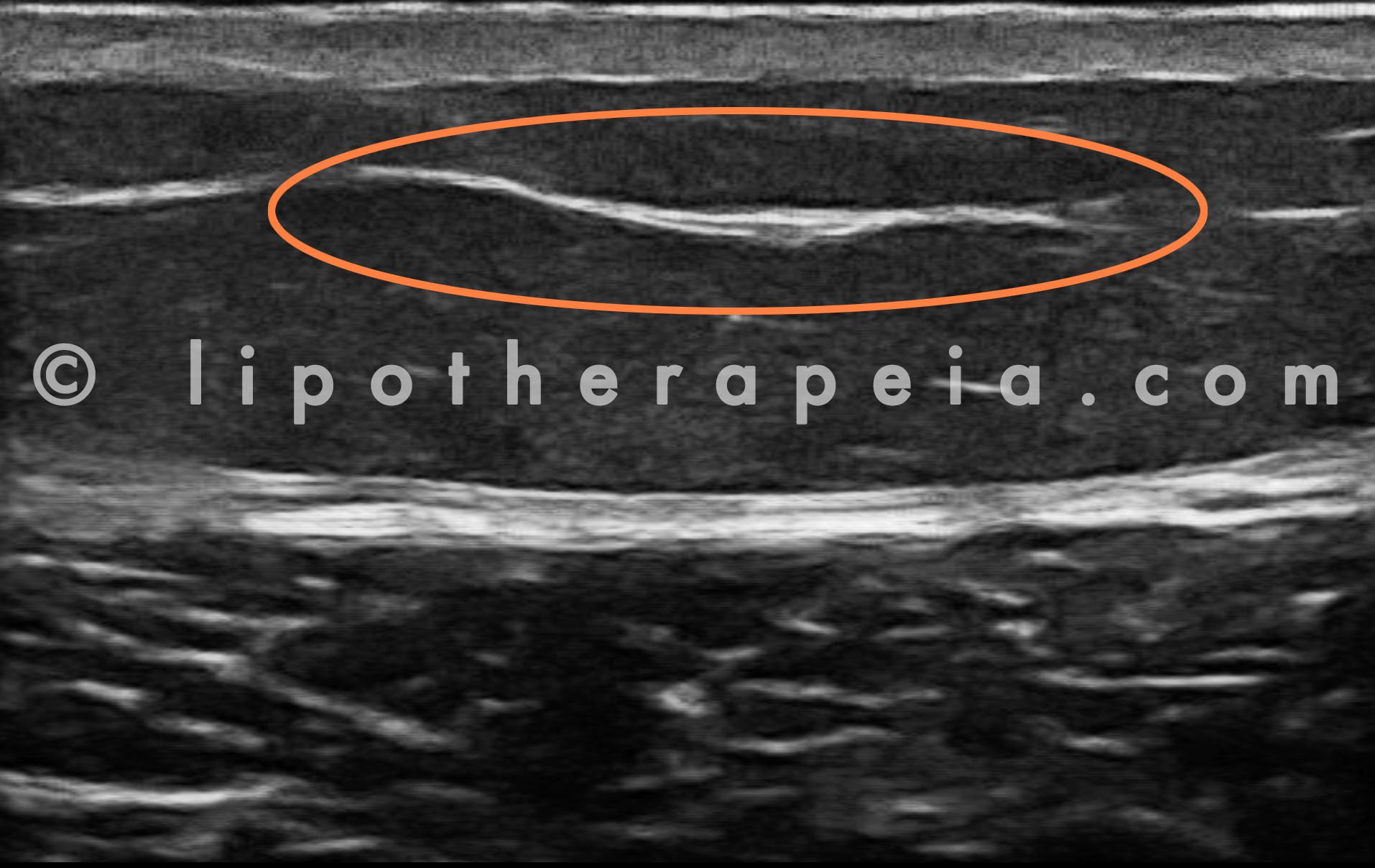
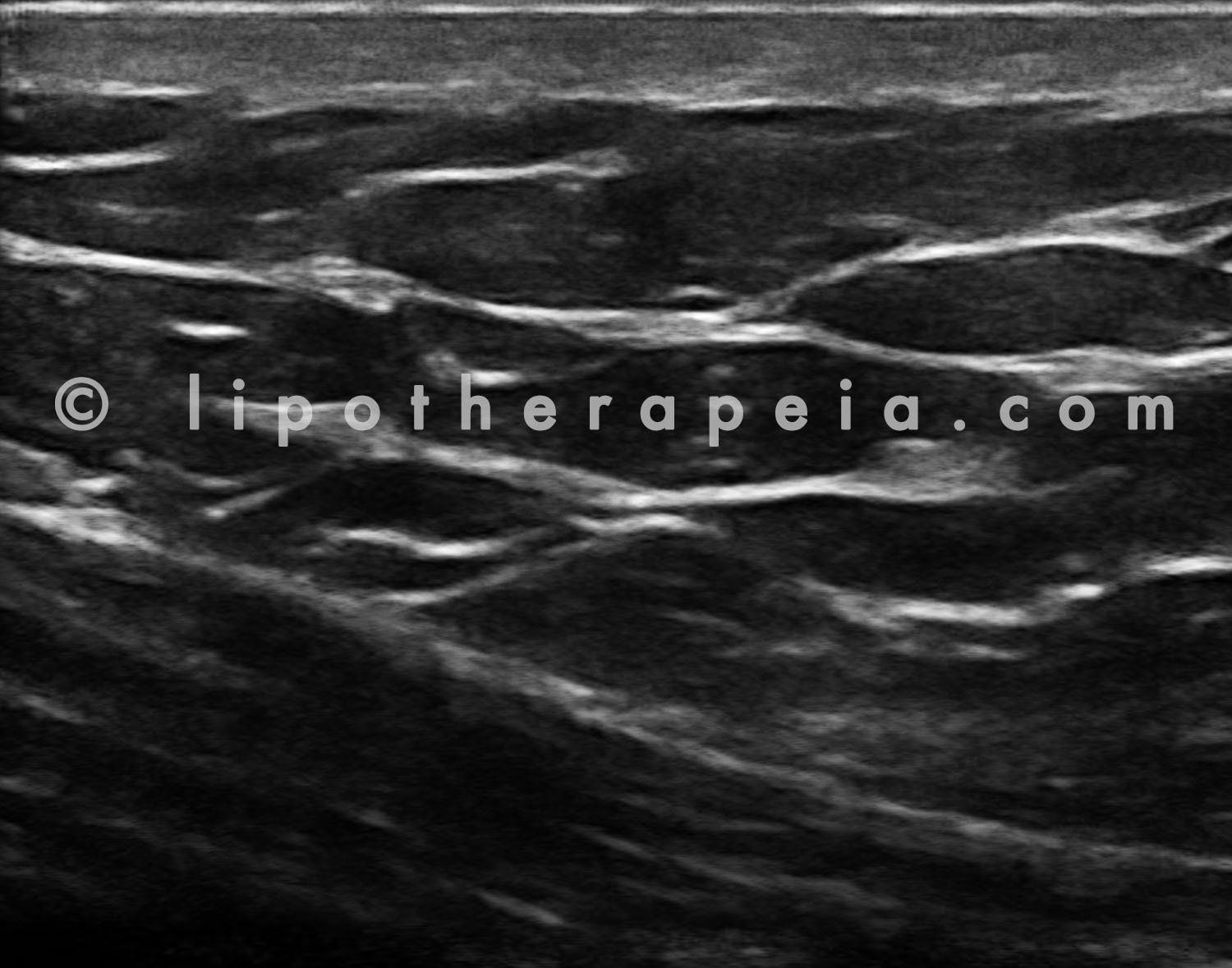
Skin ultrasound image of the lower, outer back of thigh with intermediate fascia visualised
This is an image of the lower, outer back of thigh.
Observations:
The dermis is of normal thickness for that body area
No cellulite is visible at this segment
Only one retinaculum can be observed at this spot
The superficial fascia can be observed but the intermediate one is missing (in orange)
The deep fascia is almost fused with the perimuscular fascia
At the bottom of the picture, the biceps femoris muscle can be clearly seen
Image U1 (C2)

Skin ultrasound image of the lower, outer back of thigh with deep cellulite globule
This is an image of the lower, outer back of thigh.
Observations:
The dermis is thin at this level
Some cellulite is visible
A fat globule is seen forming a cavity within the superficial fascia - this would be observed as a large cellulite bump from the outside (in orange)
The intermediate fascia is missing
The deep fascia is clearly distinguished from the perimuscular fascia
At the bottom of the picture, the biceps femoris muscle can be clearly seen
Image U1 (C7)
Loose, puffy skin and cellulite on the lower, outer back of thighs
This is a video of the lower, outer back of thigh.
Observations:
After weight loss of 10kg (1.6st) the hypodermal connective tissue at this level is extremely “spongy” and loose
No proper retinaculae are seen, just an amorphous mass of thin, weak connective tissue and multiple “islands” of fat globules
The dermis, hypodermis and subcutaneous adipose tissue look almost fused together, with no visible fascia shown between them
Upon pressure, it is compressed with the exception of large fat globules surrounded by hard connective tissue
After more than 100 clients assessed, this is the first time we saw this hypodermis / subcutaneous adipose fat appearance
This is visible from the outside as a fatty/puffy area
Image U1 (C29)
⏵ Check prices and book an expert skin ultrasound scan at our London clinic
Diagnostic ultrasound images of skin at the lower buttocks, with skin laxity

Skin ultrasound image of the lower, outer buttocks, with damaged connective tissue

Skin ultrasound image of the lower, outer buttocks, with damaged connective tissue
Loose, puffy skin and cellulite on the lower outer buttocks
These are images of the lower, outer buttocks.
Observations:
After weight loss of 10kg (1.6st) the hypodermal connective tissue at this level is extremely “spongy” and loose
No proper retinaculae are seen, just an amorphous mass of thin, weak connective tissue and multiple “islands” of fat globules (in orange)
The dermis, hypodermis and subcutaneous adipose tissue look almost fused together, with no visible fascia shown between them
After more than 100 clients assessed, this is the first time we saw this hypodermis / subcutaneous adipose fat appearance
This is visible from the outside as a fatty/puffy area
Images U1 (C15), U1 (C16), U1 (C19)
⏵ Check prices and book an expert skin ultrasound scan at our London clinic
Diagnostic ultrasound images of skin at the mid-upper buttocks, with cellulite

Skin ultrasound image of the upper buttocks, with cellulite
Skin ultrasound image of the upper buttocks, with cellulite

Skin ultrasound image of the upper buttocks, with cellulite
These are three images and video of the mid-upper buttocks.
Observations:
The dermis is a little thin for this body area
Superficial cellulite globules of various sizes can be seen
The tissue is quite fibrous, which is normal for this body area
Multiple fascia planes can be seen
The gluteus maximus muscle can be seen at some of the images
Images U1 C20), U1 (C24), U1 (C27)
⏵ Check prices and book an expert skin ultrasound scan at our London clinic